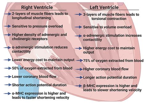
lv rv 86 | Reference (normal) values for echocardiography lv rv 86 Although the RV and LV demonstrate important differences in embryology, form, and function, they share many common characteristics when they adapt to adverse loading or . hlbdy.me. hlbdy166 has one repository available. Follow their code on GitHub.
0 · What are the echocardiographic findings of acute right
1 · Right Versus Left Ventricular Failure
2 · Right Ventricular to Left Ventricular Ratio at CT Pulmonary
3 · Right Ventricular failure
4 · Right Ventricular Function in Left Heart Disease
5 · Reference (normal) values for echocardiography
6 · Medical and Surgical Treatment of Acute Right Ventricular Failure
7 · Cardiac MRI right ventricle / left ventricle (RV/LV) volume ratio
8 · Approach to evaluation of the right ventricle in adults
9 · Anatomy, Function, and Dysfunction of the Right Ventricle:
I have a friendship invitation in e-mail, but it does not show up on frype.com? Response: There are two likely causes - 1) You are registered on frype.com with a different profile linked to that e-mail address; 2) Sender has cancelled the invitation, or the sender has been blocked from frype.com.

Normal (reference) values for echocardiography, for all measurements, according to AHA, ACC and ESC, with calculators, reviews and e-book. The right ventricular to left ventricular diameter (RV:LV) ratio measured at CT pulmonary angiogram (CTPA) has been shown to provide valuable information in patients with . Although the RV and LV demonstrate important differences in embryology, form, and function, they share many common characteristics when they adapt to adverse loading or . Evaluation of the right ventricle (RV) is a key component of the clinical assessment of many cardiovascular and pulmonary disorders. There are many ways to evaluate the RV, .
The most commonly performed and easily recognized qualitative findings are increased RV:LV size ratio and abnormal septal motion. Appearance of McConnell’s sign and . stages of RV failure. Markedly elevated CVP (central venous pressure) usually indicates RV failure in the absence of an obvious alternative explanation (e.g., severe hypervolemia, intubation with high airway pressures, abdominal compartment syndrome, pericardial tamponade). In quantitative analysis of RV size by MRI, we found that the normal RV/LV volume ratio to be 0.906–1.266. RV enlargement should be considered when this ratio is ≥1.27. This .LV dysfunction, either cytokine-induced or due to ischemia or nonischemic cardiomyopathies, induces RV dysfunction via afterload increase, and/or displacement of the interventricular septum toward the RV with subsequent impairment of RV filling (known as ventricular interdependence).
Both RV dysfunction and PHTN have independent prognostic significance. This review explores the unique anatomic and functional features of the RV and the .The RV is anatomically and functionally different from the LV, and therefore, our knowledge of LV physiopathology cannot be directly extrapolated to the right heart. The RV plays an essential role in determining symptomatic status and prognosis .
What are the echocardiographic findings of acute right
Normal (reference) values for echocardiography, for all measurements, according to AHA, ACC and ESC, with calculators, reviews and e-book. The right ventricular to left ventricular diameter (RV:LV) ratio measured at CT pulmonary angiogram (CTPA) has been shown to provide valuable information in patients with . Although the RV and LV demonstrate important differences in embryology, form, and function, they share many common characteristics when they adapt to adverse loading or . Evaluation of the right ventricle (RV) is a key component of the clinical assessment of many cardiovascular and pulmonary disorders. There are many ways to evaluate the RV, .
stages of RV failure. Markedly elevated CVP (central venous pressure) usually indicates RV failure in the absence of an obvious alternative explanation (e.g., severe . The most commonly performed and easily recognized qualitative findings are increased RV:LV size ratio and abnormal septal motion. Appearance of McConnell’s sign and .
LV dysfunction, either cytokine-induced or due to ischemia or nonischemic cardiomyopathies, induces RV dysfunction via afterload increase, and/or displacement of the interventricular .The RV is anatomically and functionally different from the LV, and therefore, our knowledge of LV physiopathology cannot be directly extrapolated to the right heart. The RV plays an essential . In quantitative analysis of RV size by MRI, we found that the normal RV/LV volume ratio to be 0.906–1.266. RV enlargement should be considered when this ratio is ≥1.27. This .
Several methods to determine RV dysfunction on computed tomographic pulmonary angiography (CTPA) have been proposed. According to the latest European Society of Cardiology (ESC) .Normal (reference) values for echocardiography, for all measurements, according to AHA, ACC and ESC, with calculators, reviews and e-book. The right ventricular to left ventricular diameter (RV:LV) ratio measured at CT pulmonary angiogram (CTPA) has been shown to provide valuable information in patients with .
Although the RV and LV demonstrate important differences in embryology, form, and function, they share many common characteristics when they adapt to adverse loading or . Evaluation of the right ventricle (RV) is a key component of the clinical assessment of many cardiovascular and pulmonary disorders. There are many ways to evaluate the RV, .
Right Versus Left Ventricular Failure
stages of RV failure. Markedly elevated CVP (central venous pressure) usually indicates RV failure in the absence of an obvious alternative explanation (e.g., severe .
The most commonly performed and easily recognized qualitative findings are increased RV:LV size ratio and abnormal septal motion. Appearance of McConnell’s sign and .
LV dysfunction, either cytokine-induced or due to ischemia or nonischemic cardiomyopathies, induces RV dysfunction via afterload increase, and/or displacement of the interventricular .The RV is anatomically and functionally different from the LV, and therefore, our knowledge of LV physiopathology cannot be directly extrapolated to the right heart. The RV plays an essential . In quantitative analysis of RV size by MRI, we found that the normal RV/LV volume ratio to be 0.906–1.266. RV enlargement should be considered when this ratio is ≥1.27. This .

Right Ventricular to Left Ventricular Ratio at CT Pulmonary
Right Ventricular failure
When it finds a Sableye trying to catch a Carbink, Gabite becomes furiously angry and attacks the Sableye. ultra-moon: It sheds its skin and gradually grows larger. Its scales can be ground into a powder and used as raw materials for traditional medicine.
lv rv 86|Reference (normal) values for echocardiography


